Methods and approaches
Our focus is the quantitative analysis of cell biological mechanisms in heterogeneous cell models, in particular using microscopy-based methods such as High Content Screening (HCS). This includes the development of novel assays and the extensive automation of microscopy as well as image and data analysis. With this approach, we can record multiple cellular parameters in thousands of cells simultaneously and objectively. These can be e.g. the number of cells, cell size, relative position, morphological properties, immunofluorescence intensities of marker proteins, number of transcripts per cell, or the subcellular localization.
In the context of pain research, we use HCS microscopy mainly to quantify signal transduction in primary nociceptive neurons, associated glial cells of the dorsal root ganglia, and iPS-derived nociceptors. For this purpose, we have developed novel methods to determine the activity of the crucial signaling cascade component, PKA-II, directly in neurons without the need of reporter systems.
In collaborations, we also apply our approaches to cell biological mechanisms in other cell models. For example, we provide our expertise regarding high throughput microscopy to colleagues investigating cellular stress responses or DNA repair mechanisms in patient fibroblasts and cancer cell models. Please contact us if you would like to apply HCS to your cell model as well.
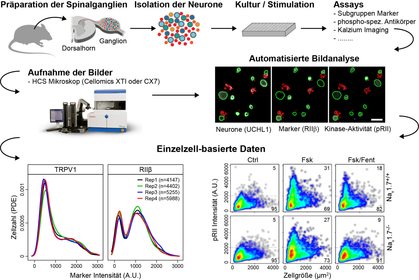
After enzymatic digestion of dorsal root ganglia, the isolated cells are cultured in mutiwell plates, stimulated and analysed either live or fixed using HCS microscopy (Cellomics XTI or CX7). Various functional assays allow quantification of the cellular response in marker-defined subsets of sensory neurons (Figure by Jörg Isensee).

To express channelrhodopsin 2 (ChR2, bottom left, green), nociceptive neurons were transduced with adeno-associated viruses (AAV-PHP.S). The optogenetically excitable cells can then be stimulated using OptoPlate 96 (Bugaj et al., 2019) and analyzed using HCS microscopy (Figure by Jörg Isensee).
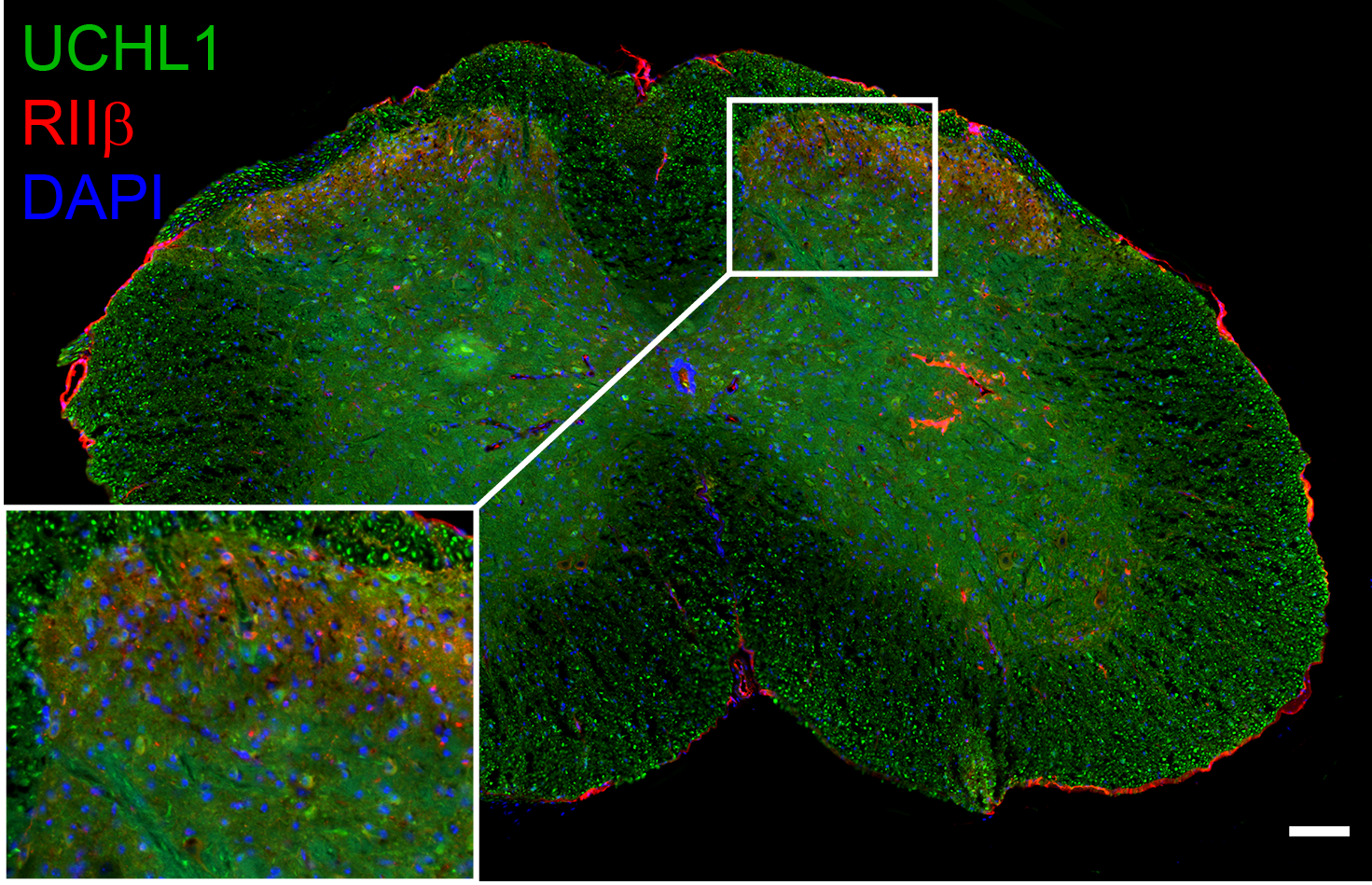
Frozen section of the mouse spinal cord. The neuronal marker UCHL1 and the PKA subunit RIIβ were labeled by immunocytochemistry, the nuclei were stained with DAPI. RIIβ was detected in the outer laminae of the dorsal horn, where the central termini of nociceptive neurons are located (figure by Jörg Isensee).
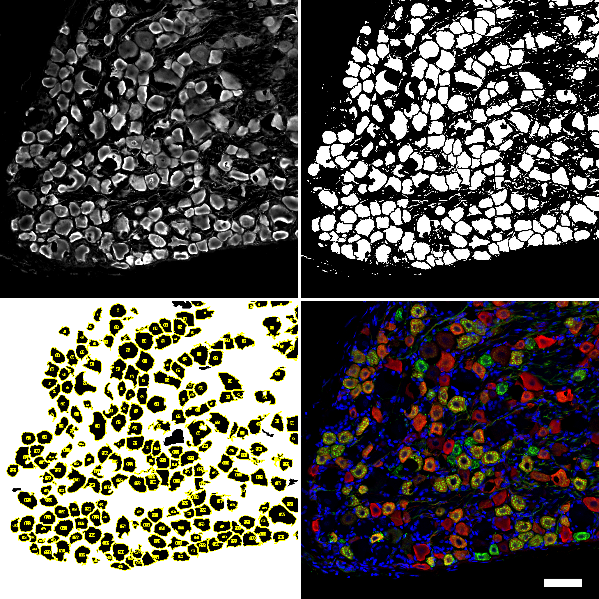

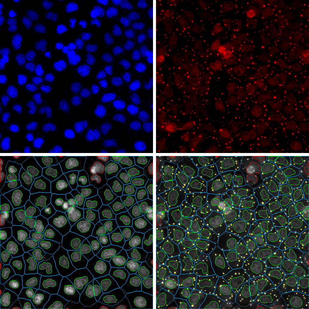
Confocal images of frozen sections of lumbar dorsal root ganglia after immunofluorescence staining (10 µM thick, scale = 50 µM). The neuronal marker UCHL1 (grey) was used to create an image mask (bottom left), which allows quantification of the expression of subgroup-specific markers RIIβ (red) and IB4 (green) (figure by Jörg Isensee).
Penk mRNA was detected by fluorescent in situ hybridisation (FISH), the neuronal marker UCHL1 (green) by immunofluorescence and the cell nuclei by Hoechst 34580.

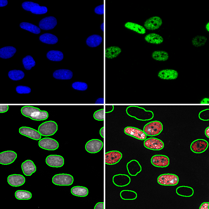
To form stress granules (top right, red), HeLa cells were treated with 0.5 mM sodium arsenite for 1 h (figure by Christian Kähler / Jörg Isensee).
The cell bodies of iPS-derived nociceptors were stained with fluorescent Nissel (green), the neurites by immunofluorescence against the PKA subunit RIIβ, the nuclei with Hoechst 34580 (figure by Andreea Belu).
Primary dermal fibroblasts were treated with hydroxyurea (20 mM) for 1 h and fixed after 48 h (figure by Ron Jachimowicz / Jörg Isensee).
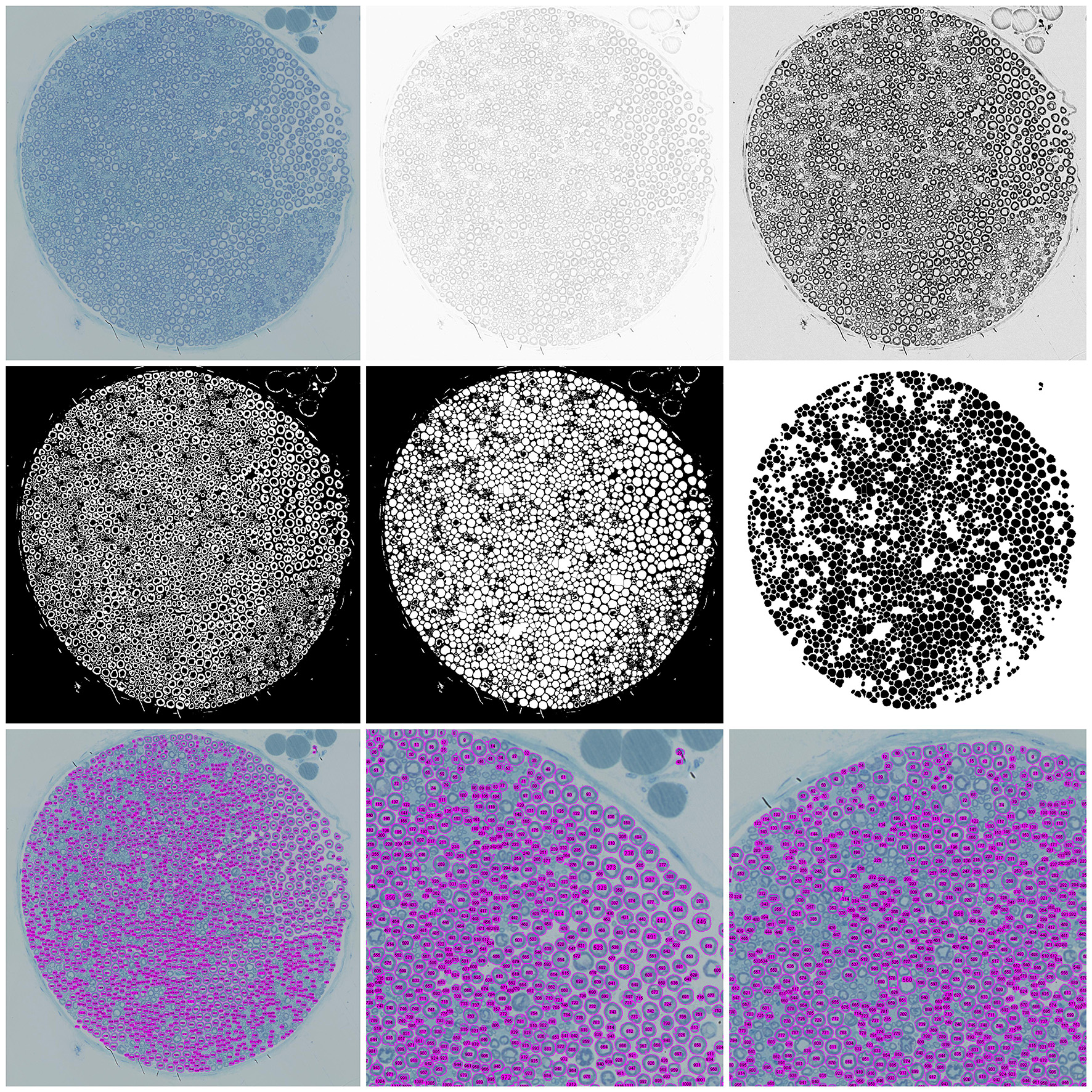
Phase-contrast images of frozen sections of the sciatic nerve were used to create a mask that allows quantification of the number and size of nerve fibres (illustration by Ilja Bobylev / Jörg Isensee).